- 77 Posts
- 60 Comments

 1·1 year ago
It’s not, but it’s very painful.

 2·1 year ago
No they don’t all calcify.
The tumor here is growing from the bone and destroying it. The normal bone cells are trying to repair by making more bone. If faster the tumor destroys and grows, the less smooth the repaired bone can look.

 2·1 year ago
Yeah they can decompress by removing part of the back of the skull so there’s more space.

 2·1 year ago
It’s called periosteal reaction. Here’s a few examples I posted already: one, two.

 2·1 year ago
Hi. Try to match the outline of the humerus to that on the radiograph. Then the big bright mass to the right of the humerus (going by the picture) is the cancer. Now see if you can make out the rough outline of it on the radiograph.
The cloudiness you’re seeing is calcification within the tumor. Specifically, the outward “tendrils” (as someone else called it) exploding out from the margin of the humerus is really aggressive periosteal reaction, which I’ve posted a few examples before already: one, two.
I think while the general communities have made it, a lot of niche communities failed to attract enough population to keep on generating more content. As an example, just search for the “Imaginary” series of landscape art communities on the Fediverse (eg. ImaginaryVistas). Many of them don’t have any recent posts or 1 post per days or weeks. That’s not enough to keep people invested. Even the largest digital art community is still mostly carried by 1 person.
The Lord said that the “VCR shall be saved” with the knife technique, but in the following paragraph, it was not the VCR that was saved, but the man that was saved. The VCR was not saved!

 1·1 year ago
Developmental delay, seizures, cerebral palsy, autism, depending on severity.

 2·1 year ago
It’s usually barium sulfate, which can have a more viscous consistency that makes it go down a little slower and allows it to stick to things to outline them. But yes, regular iodine contrast can also be used.

 2·1 year ago
Yep. They’re in the middle of swallowing some liquid contrast. Take the picture 1 second later and you miss the shot.

 11·1 year ago
It looks like it could fall over with any gust of wind and kill someone.

 71·1 year ago
Should the instances that responded to you be refederrated? I’m pretty sure I saw some of them on lemmy.world’s block list. I think it would be sad for these small servers to not realize they are, in fact, not connected to the greater fediverse. On the other hand, if you’re an admin, and you don’t know what you’re doing to the point of not knowing your server was infected by hundreds of thousands of bots, maybe it’s too dangerous to refed.

 3·1 year ago
I think these are valid concerns, once again, and must be discussed and hashed out so that everyone is on the same page.
The medical field has a long history of sharing cases for education, and I would say radiology definitely is one of the fields where this is done more often than not, due to the complexity of cases, use of imaging, which is a format that makes it easier to share. I would also say that radiology is quicker to embrace new technologies, including use of the internet and social media, due to reliance on technology to begin with. For example, you can find on twitter, of all places, many case presentations and discussions, many by prominent leaders in the field. Almost all the major radiology societies have some case series for perusal, there’s Radiopaedia as I already mentioned, and the largest community I’m aware of is r/radiology, which I modeled this community after.
I understand there’s a slippery slope between posting educational content and entertainment content. I intend to firmly keep us away from the entertainment side. While I do not want this community to fall into a somber tone, I will not tolerate posts or comments intended to mock or demean a case/patient.
In regards to data harvesting - interesting thought. I had to dig a little bit. There’s definitely been some publications that deal with large medical imaging datasets wherein AI was able to use the high resolution imaging to identify patient features, including 3D reconstruction of the face from the data. The images posted here are: 1) regular .jpgs and not high resolution files like dicom, 2) limited to a couple of images when a full CT can be in the hundreds, and 3) been through photoshop, powerpoint, possibly multiple saves though jpg with degradation each time, or they’re crappy phone camera images of a medical image. I can’t say the risk is absolutely 0, but it’s pretty close - by staying within medical privacy laws and avoiding super rare presentations of things.
However, your concerns have been my concerns too, so I have also reached out to the .world admins for their thoughts. Perhaps they don’t feel comfortable hosting this type of content, or would like to see some other assurances. I will find out and proceed from there.

 4·1 year ago
Yes that’s correct, it’s the levamisole that presented as demyelination in this case.
Cocaine by itself can also cause problems, which I would categorize in 3 ways:
-
Sudden-onset issues that are potentially life-threatening. Cocaine is a stimulant, which can raise blood pressure, heart rate, etc. Those effects can cause ischemic stroke, hemorrhagic stroke, rupture of pre-existing aneurysms (with subsequent intracranial hemorrhage), and seizures, all of which can show up on brain imaging.
-
Long-term issues. These are insidious, subtle changes from repeated use of cocaine, which will damage the brain through neurotoxicity + the long-term results of an accumulation of events from #1.
-
Complications from cocaine use. I would put levamisole in this category. For other drugs that are more commonly injected (rather than snorted or smoked), brain infections from use of dirty needles would also go into this category.
-

 11·1 year ago
Thank you for this comment, because it brings up some very important issues, which I hope this reply addresses.
The biggest issue is the matter of patient confidentiality. This is of utmost concern in an online medical community, especially one wherein clinical vignettes are presented. I take extreme care to avoid including any information that can narrow down to a patient, thus breaching confidentiality. Similarly, I expect anyone else commenting or posting here to follow this rule, Rule 4, which was created not just for internet etiquette but literally to prevent illegal breaches of confidentiality. With regards to consent - this is not required for publishing de-identified information, or sites like Radiopaedia with their thousands of cases would not exist. With regards to patient confidential information that cannot be shared - this not only includes the obvious ones such as patient age, DOB, dates of events, addresses, etc etc, but also vaguer information. For example, there are cases that I would never present here online because the disease is so incredibly rare that the disease itself becomes a patient identifier. These types of cases I would formally publish in the literature if need be. For this particular case, I do not think the information presented breaks these rules (or I would not have posted). Cocaine use is fairly common among the demographic in question, and being found in the shower is not that uncommon, although dramatic.
Second, to address the following:
The title is like your a friend but the text is from a medical professional.
This is something that I have been struggling with. This community is growing at an exceptional rate, and visitors seem to be overwhelmingly from a non-medical background. There are comments that frankly say they do not understand certain things, and other comments and questions imply a lack of experience with looking at imaging studies. I have been vacillating between using terminology and sentences that laypeople can understand versus maintaining medical terminology. I think this is why you think I am writing about a friend in the title, but the body of the post is more medically-oriented. For this reason, I have changed the title - it did not need to be so dramatic. In the future, I will be more careful with my wording.
Please let me know if this addresses your concerns. I would also love to hear more input regarding point #2. Should I continue to word cases as if talking to other medical professionals or include more basic terminology so that the general public can understand? The purpose of these cases, and this community in general, is to be an educational resource in terms of what Radiology is and does.

 4·1 year ago
This complication of cocaine use generally manifests as a single episode of demyelination that recovers well with treatment. Of course, the long term depends on how often the cocaine abuse happens.

 2·1 year ago
Oh good to know. I guess I only really caught reddit downtimes in the past.

 82·1 year ago
I like how it looks exactly like reddit’s status page, especially considering they just released old.lemmy.world.

 1·1 year ago
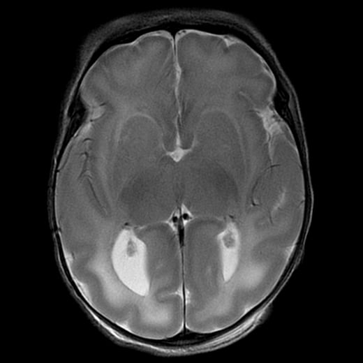
Just a standard T2 sequence.
FLAIR would have dark CSF, since that’s what FLAIR is designed to suppress - CSF.
MPRAGE is a T1 sequence that’s usually done with contrast.


Much further back… Abraham Lincoln was a Republican. The party was founded on anti- slavery. The current party has nothing to do with the original party.